FDPNP3: Artificial Intelligence under the Microscope (Part 2)
Preface: I want to initiate a comprehensive series, “Fundamentals of Digital Pathology for Novice Pathologists (FDPNP)”. This endeavor aims to disseminate knowledge about the utilization of computer vision in the realm of modern pathology to pathologists who want to know more about digital pathology.
Introduction
In part 1, we briefly decoded the complicated relationships between AI-related concepts. In this part, we use that knowledge to clarify where AI stands in digital pathology.
Having gained a general understanding of how these buzzwords interrelate, let’s delve into the subfields of AI that are utilized in digital pathology.
The bullet point of this mini-review:
In digital pathology, the main AI applications involve computer vision, machine learning, and natural language processing. These are anticipated to evolve in a more sophisticated manner in the future.
Computer vision
CV is a field that involves sophisticated algorithms capable of managing and processing visual data such as images and videos. These algorithms transform this visual data into valuable information relevant to the task at hand.
Computer Vision is the core application of AI in digital pathology. Almost all pathology-related tasks involve this subfield
The main idea of using CV in digital pathology:
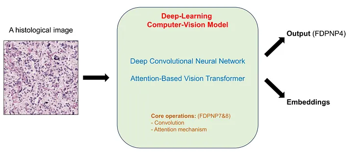 The main idea of using computer vision in digital pathology - Image by Author
Note: the “output” and “embedding” parts seem ambiguous. We will clarify “output” in Part 3(FDPNP4) and “embedding” in the subsequent blogs when we investigate the internal operation of these models.
CV largely relies on Deep Learning (DL) models. Currently, there are two primary types of DL models used in CV: Deep Convolutional Neural Networks (DCNNs) and Vision Transformers (ViTs). Each category encompasses numerous DL models, which are beyond the scope of this mini-review. These two types are named after their fundamental operations: convolution and the attention mechanism. We will delve into these two operations in FDPNP7 and FDPNP8.
Machine Learning (ML)
The concept of ML was explained in part 1. In this part, I specifically refer to the classic ML models, which require structured input data. Recall that structured data is one that can be organized into a data table.
ML involves the extraction of histological features, which can be performed by either human pathologists or computer vision (CV) models. Once these morphological characteristics are processed by human observation or CV techniques, they are transformed into structured data. This data may take the form of categories, numerical variables (when processed by the human eye), or a feature representation vector (when processed by CV). These features can subsequently be utilized by Machine Learning models to execute the task at hand.
For instance, metrics such as mitotic count, tumor-infiltrating lymphocytes, and the tumor-stroma ratio are estimated by humans and can be either categorical or numerical. An intermediate-layer vector, generated by a trained risk-stratifying CV model, can serve as a histopathological representation of risk. This can then be used as an input for ML algorithms.
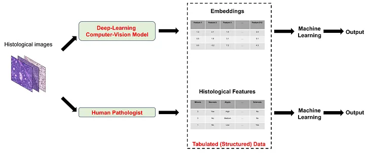 The ways to apply machine learning in interpreting histological images - Image by Author
Natural Language Processing (NLP)
Much like CV with visual data, NLP is a field that involves sophisticated algorithms capable of managing and processing textual data such as letters or articles.
In digital pathology, NLP is commonly employed for tasks such as text-to-image generation (for instance, generating images from caption prompts) or image-to-text generation (like automated reporting). The primary objective of these tasks is to lessen the workload of pathologists and to contribute to educational efforts. However, this technology is still in its infancy and not yet ready for practical implementation. Nonetheless, with continuous research and development, we can anticipate more practical applications in the future.
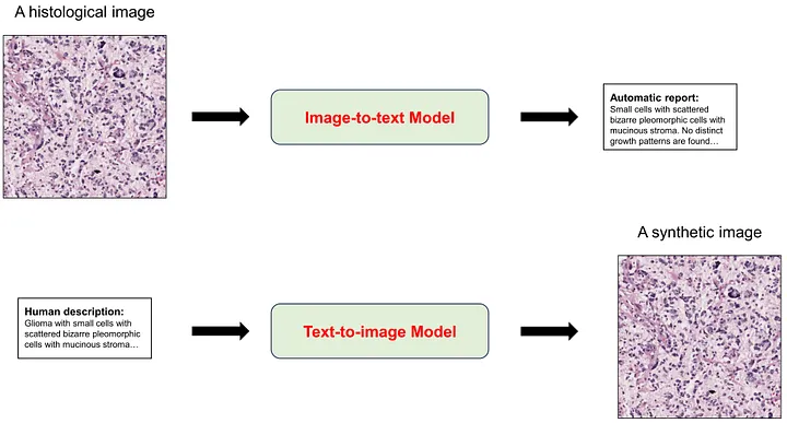 Using natural language processing in digital pathology - Image by Author
Conclusion
Various subdomains of AI find their application in pathology, with computer vision being the fundamental method. In our upcoming blog, we will delve into the category of pathology-related AI tasks.
For those who may be interested, I hope you enjoy this mini-review.